Deadlines
Abstract submission
Acceptance notice
Early registration
If you need financial support to attend the 29th CSBMM, submit your abstract before July 16th.
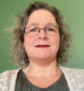
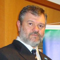
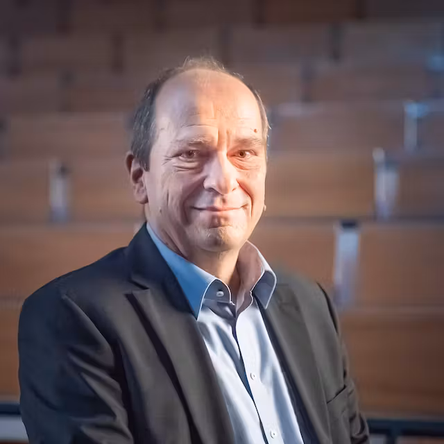
Organization
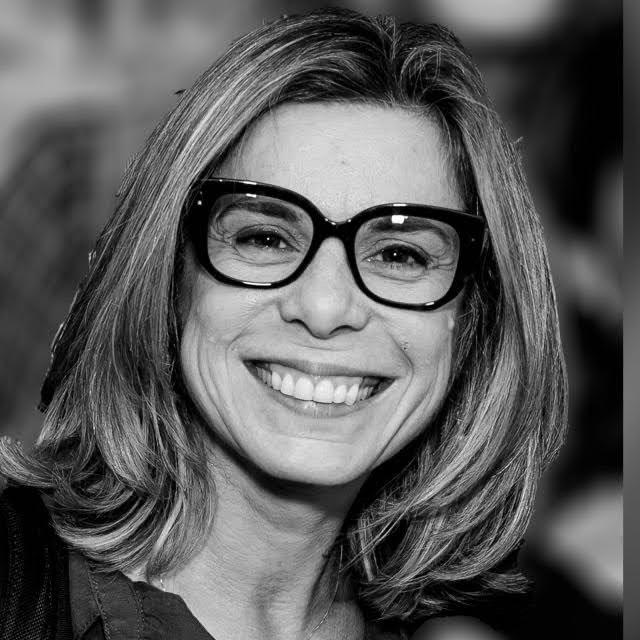
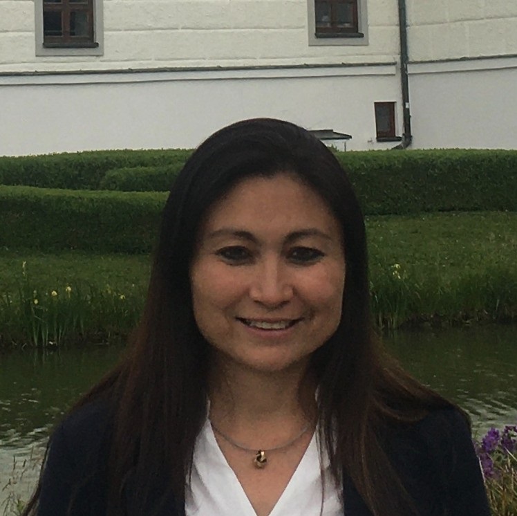
Giovanna Machado
CSBMM Chair
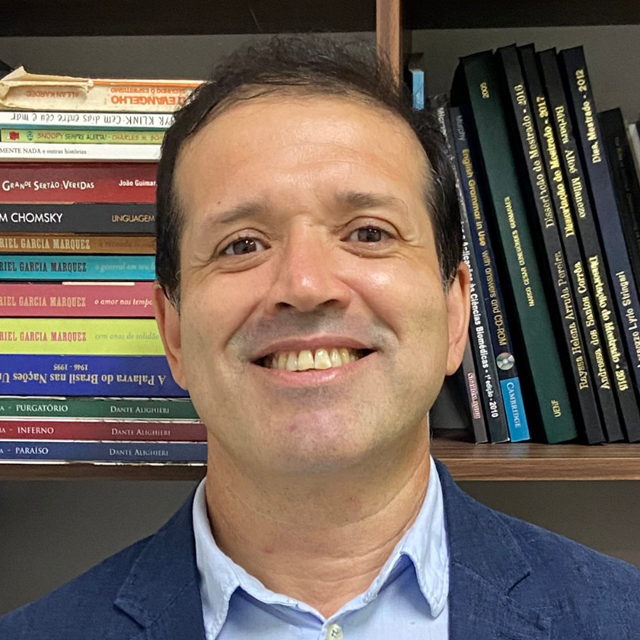
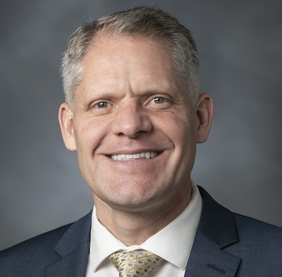
Marco Guimarães
SBMM President
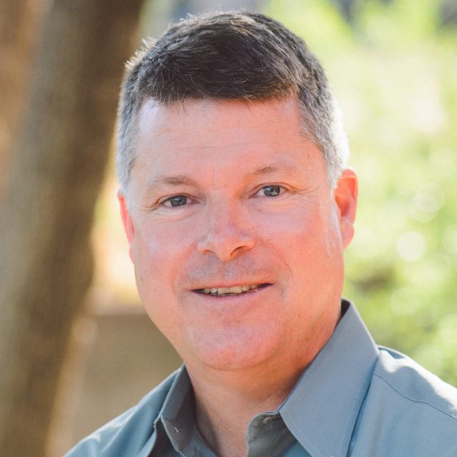
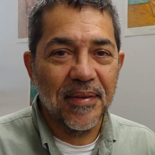
Paul Kotula
CIASEM President
About
We are pleased to present the 29th Congress of the Brazilian Society of Microscopy and Microanalysis and the XVII Interamerican Congress on Microscopy. This is certainly the most important scientific activity in Brazil and Latin America.
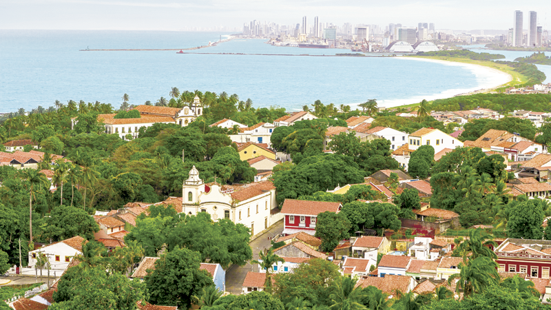
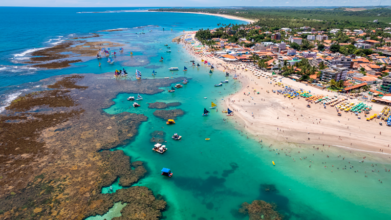
Olinda is a very exciting place and has become a hub for artists and bohemian types and the twisting streets are full of galleries, museums, workshops and quaint churches. The area was designated a UNESCO World Heritage site in 1982.
Presentation
Dear microscopists,
Welcome to the 17th Interamerican Congress on Microscopy (17th CIASEM) and the 29th Meeting of the Brazilian Society for Microscopy and Microanalysis (29th CSBMM), which will take place in Olinda, Pernambuco, from November 7th to 10th, 2023. This is a scientific event, organized by the Interamerican Committee of Microscopy Societies (CIASEM) and the Brazilian Society for Microscopy and Microanalysis. The purpose of the CIASEM shall be to increase and diffuse, for scientific and educational goals, the science, practice, and instrumentation of microscopy on the American continent.
It is the largest microscopy and microanalysis event in Latin America, with direct contributions to the advancement of science, technology, and innovation in the Southern Cone. The event promotes an environment for disseminating the main research findings in the thematic areas and discussing the contemporary challenges of research on professional training in microscopy and microanalysis. This includes exploring all practical dimensions of systematized implementation, such as the use of artificial intelligence for pattern recognition, to encourage scientific demands across different microscopy and microanalysis approaches.
The main contributions of the CIASEM/CSBMM up to the present day are: scientific outreach; recognition and promotion of microscopy and microanalysis as essential tools for research and technological development, by incorporating their results as indicators of research quality and providing theoretical and practical support for their use; and the encouragement of discussions on the theme of the event and promotion of professional training in microscopy and microanalysis.
The target audience of the CISEM/CSBMM is undergraduate and graduate students, researchers, university professors, companies, and professionals from the private sector who are interested in the field of microscopy and microanalysis.
This year’s meeting will be particularly special, as it will be held jointly with CIASEM, currently under the presidency of Dr. Paul Kotula (Sandia National Laboratories, USA) and CSBMM, under the presidency of Dr. Marco Guimarães (Federal University of Espirito Santo, Brazil), reinforcing the international character of the Congress, which is already the largest microscopy event in Latin America.
See you in Olinda, Brazil.
Regards,
CIASEM and SBMM boards
Confirmed speakers
Claire M. Brown (Canada)
McGill University
Daniel Ugarte (Brazil)
Univ. Estadual de Campinas (UNICAMP)
Dr. M. Grace Burke (USA)
Idaho National Laboratory
Joachim Mayer (Germany)
RWTH Aachen University
Grant Jensen (USA)
Brigham University
Hellen Ishikawa-Ankerhold (Germany)
Ludwig Maximilians Universität
Marcos Farina (Brazil)
Federal University of Rio de Janeiro
Social Programs

Opening Cocktail
Celebration
Tours
Awards and Competitions
Poster awards
Presentation awards
Pitch elevator
Hosted by

Sponsored


Supported by
Registration
| Until November 7th | |
|---|---|
| Professionals* | BRL 685,00 |
| Graduate students and postdocs* | BRL 365,00 |
| Undergraduate students and technicians* | BRL 250,00 |
| Companies delegates** | BRL 660,00 |
*SBMM members will be free from the 2022 and 2023 membership
*First-time SBMM members will be free from 2023 membership
**50% off for delegates from companies associated with SBMM
Mediante o pagamento de inscrição:
*Associados SBMM vão estar isentos das anuidades 2022 e 2023
*Novos membros associadaos SBMM estarão isentos da anuidade 2023
**50% de desconto para delegados de empresas associadas à SBMM
First-time SBMM members, click here.
Pre-event agenda
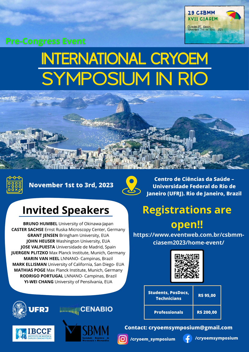
International CryoEM Symposium in Rio
Welcome to the International CryoEM Symposium in Rio de Janeiro, Brazil. This exciting event is set to take place from November 1st to 3rd, 2023 and promises to be an incredible opportunity to explore the latest developments in cryo-electron microscopy. We are thrilled to bring together some of the most renowned researchers and experts in the field of cryoEM from around the world to share their insights, discoveries, and knowledge. The symposium is all set to explore different areas of cryomicroscopy, including quick freeze of biological samples, freeze fracture, freeze-etching, cryo-scanning microscopy, freeze-substitution, single particle analysis, cryolamella preparation and cryo-electron tomography. This Symposium was idealized to celebrate the inauguration of the Cryo Microscopy Sector of the National Center for Structural Biology and Bioimaging (CENABIO), that is localized at the Federal University of Rio de Janeiro (UFRJ). The CENABIO already count with an Advanced Microscopy Facility and the expansion of this, with implementation of a dedicated cryomicroscopy sector, is of great importance for Brazilian science.
Organization: SBMM; UFRJ and CENABIO
Date: 1st to 3rd November 2023
Location: Centro Nacional de Biologia Estrutural e Bioimagem, Unidade 3 - Microscopia Avançada (CENABIO 3 - UFRJ).
Universidade Federal do Rio de Janeiro UFRJ
Unidade de Microscopia Avançada.
Av. Carlos Chagas Filho, 373 - Centro de Ciências da Saúde - CCS - Bloco M - CENABIO
Cidade Universitária - Rio de Janeiro, RJ
Contact: sbmm@sbmm.org.br
More details: https://www.instagram.com/cryoem_symposium/
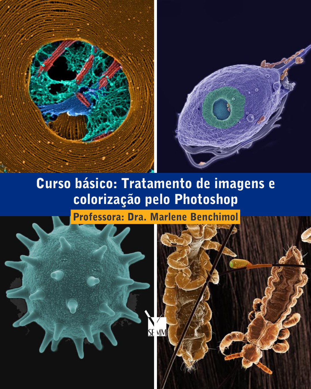
Tratamento de imagens e colorização pelo Photoshop - curso básico: demonstrativo e prático
Data: Segunda-feira 06 de novembro de 2023
Horário: 13:30h às 17 horas (on-line)
Professora: Dra. Marlene Benchimol (marlenebenchimol@gmail.com)
10 vagas - online
Requisitos: Obrigatório ter o Photoshop instalado, preferencialmente o CS6 Ementa: O que ilegal e legal no tratamento de imagens de microscopia para teses e publicações como melhorar a qualidade de imagens microscópicas para publicação dentro da legalidade. Como colorizar imagens biológicas de microscopia e Treinamento prático.
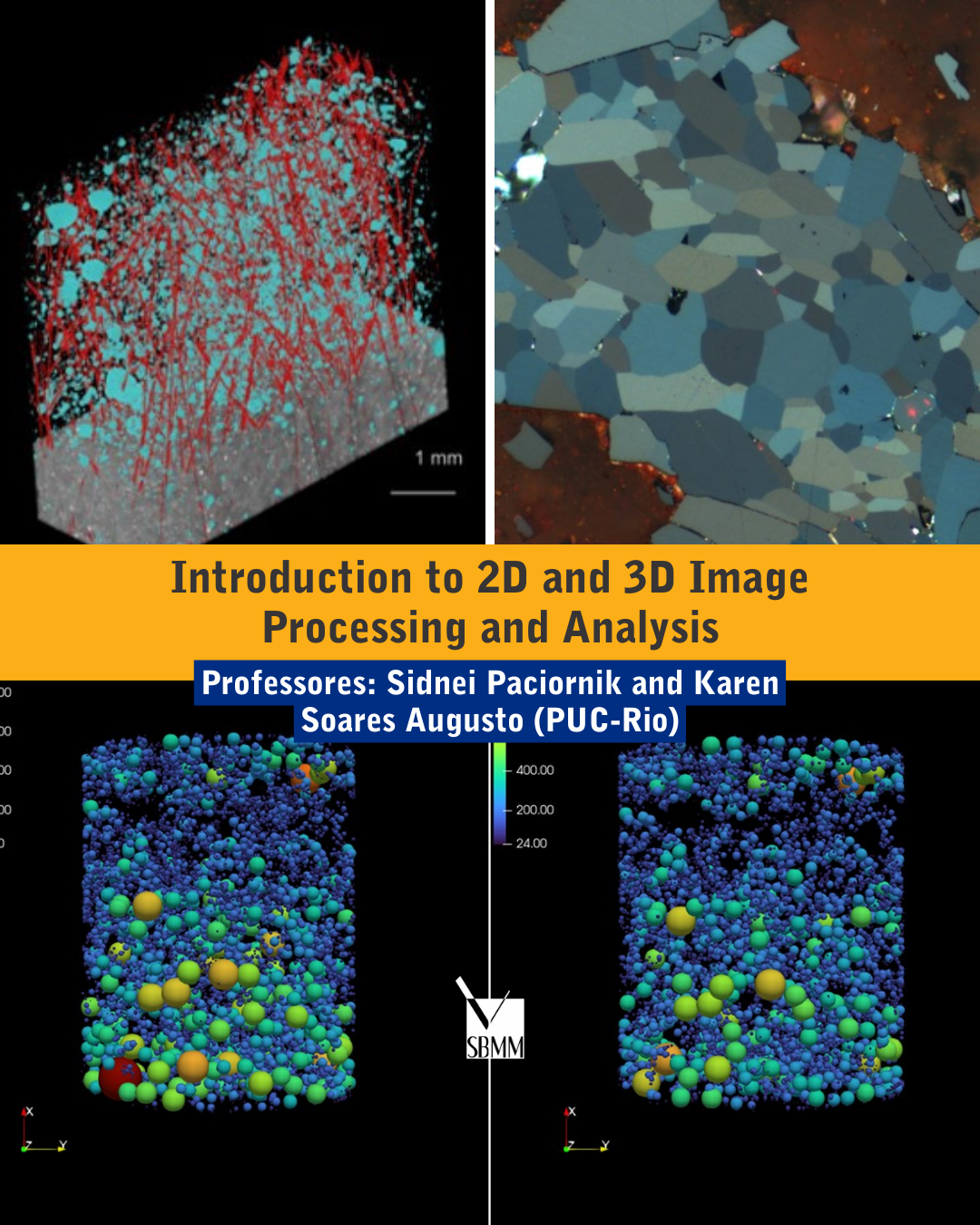
Introduction to 2D and 3D Image Processing and Analysis
This mini-course presents the principles of image processing and analysis methodology applied to 2D and 3D images obtained by different microscopy techniques. The traditional sequence - acquisition, pre-processing, segmentation, post-processing, measurements, and classification - will be presented. New features based on AI and Deep Learning will also be discussed. No previous knowledge is needed. The short course is appropriate for both materials and biology researchers.
Requirement: bring your computers
Date: November 6th (Monday) between 9AM-12PM and 1PM-5PM and November 7th (Tuesday) between 9AM to 12PM.
Location: FPE (more details soon)
Professors: Sidnei Paciornik (PUC-Rio) and Karen Soares Augusto (PUC-Rio)
Contact: sidnei@puc-rio.br and saugustokaren@gmail.com
Vacancies: 30
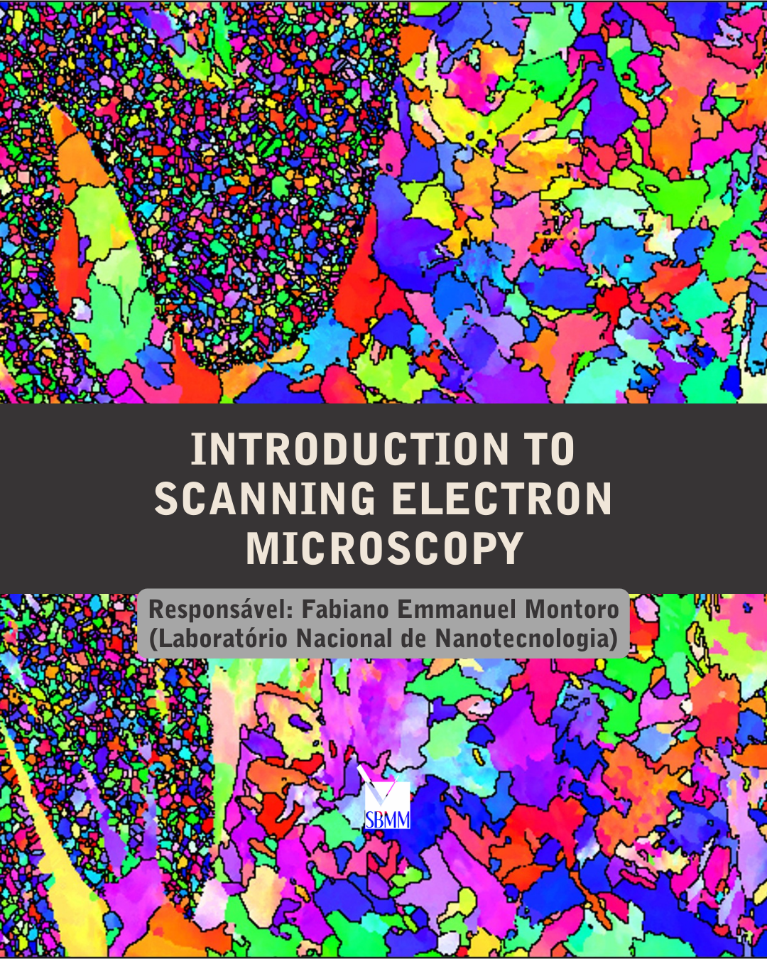
Introduction to Scanning Electron Microscopy
Date/hour: November 6th (Monday) between 9AM to 5PM and November 7th (Tuesday) between 9AM to 12PM.
Location: LACMAT-LAMICRO/CETENE (Recife/PE).
Professors: Fabiano Emmanuel Montoro (LNNano – Laboratório Nacional de Nanotecnologia)
Vacancies: 20 - in person (Target Audience: Undergraduate and postgraduate students and technicians)
Objectives: Theoretical and experimental (demo) basic course of scanning electron microscopy (SEM), offering introductory knowledge about the technique and its applications.
Duration: 10 hours.
Contact: fabiano.montoro@lnnano.cnpem.br
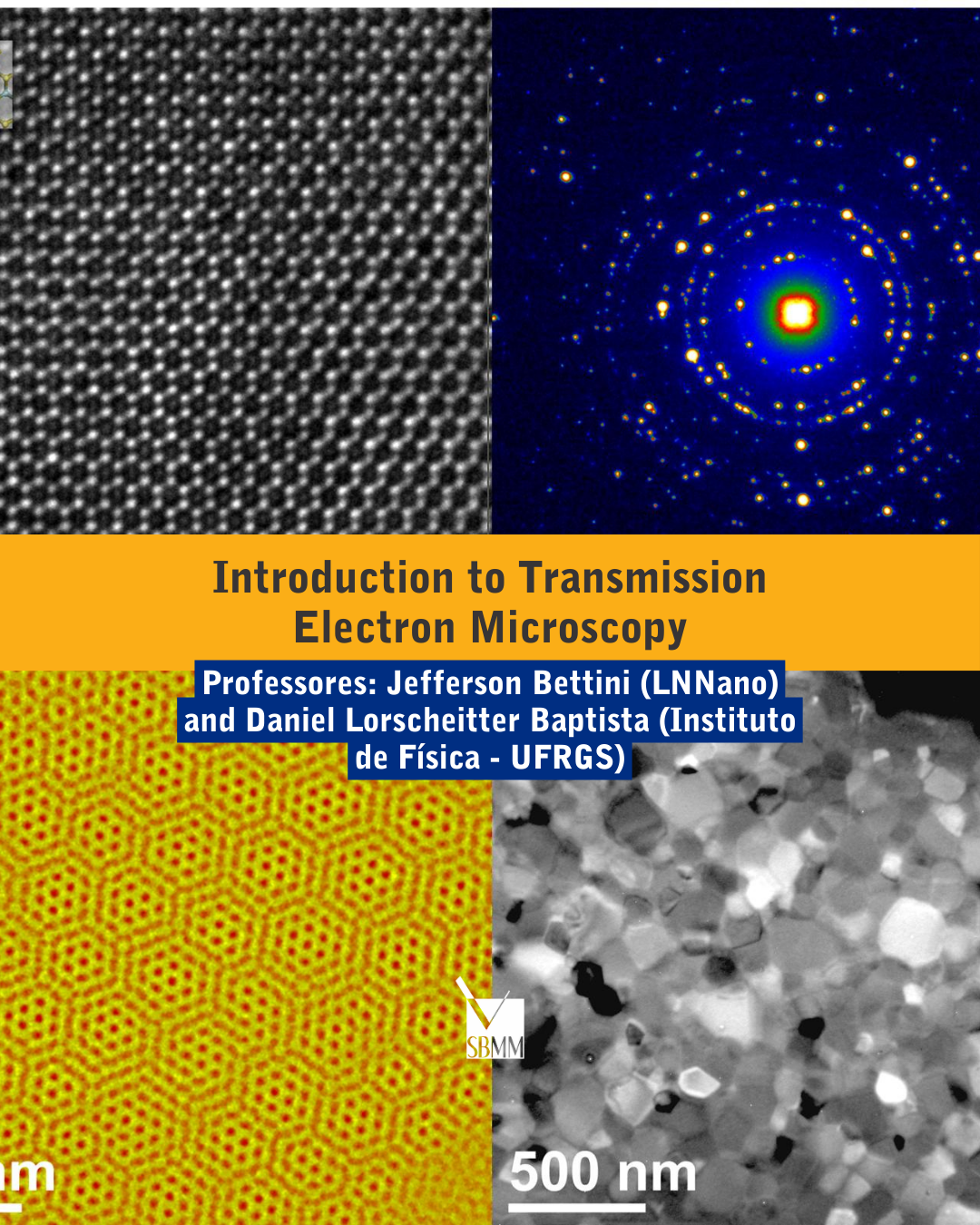
Introduction to Transmission Electron Microscopy
Date/hour: November 6th (Monday) between 9AM to 5PM and November 7th (Tuesday) between 9AM to 12PM.
Location: LACMAT-LAMICRO/CETENE (Recife/PE).
Professors: Jefferson Bettini (LNNano – Laboratório Nacional de Nanotecnologia) and Daniel Lorscheitter Baptista (Instituto de Física - UFRGS)
Vacancies: 20 - in person (Target Audience: Undergraduate and postgraduate students and technicians)
Objectives: Theoretical and experimental (demo) basic course of Transmission Electron Microscopy (TEM). This course will provide knowledge to plan their experiments, parameters to be used to obtain their results in the microscopy and interpretation of the data set.
Duration: 11 hours.
Contacts: jefferson.bettini@lnnano.cnpem.br and dbaptista@gmail.com
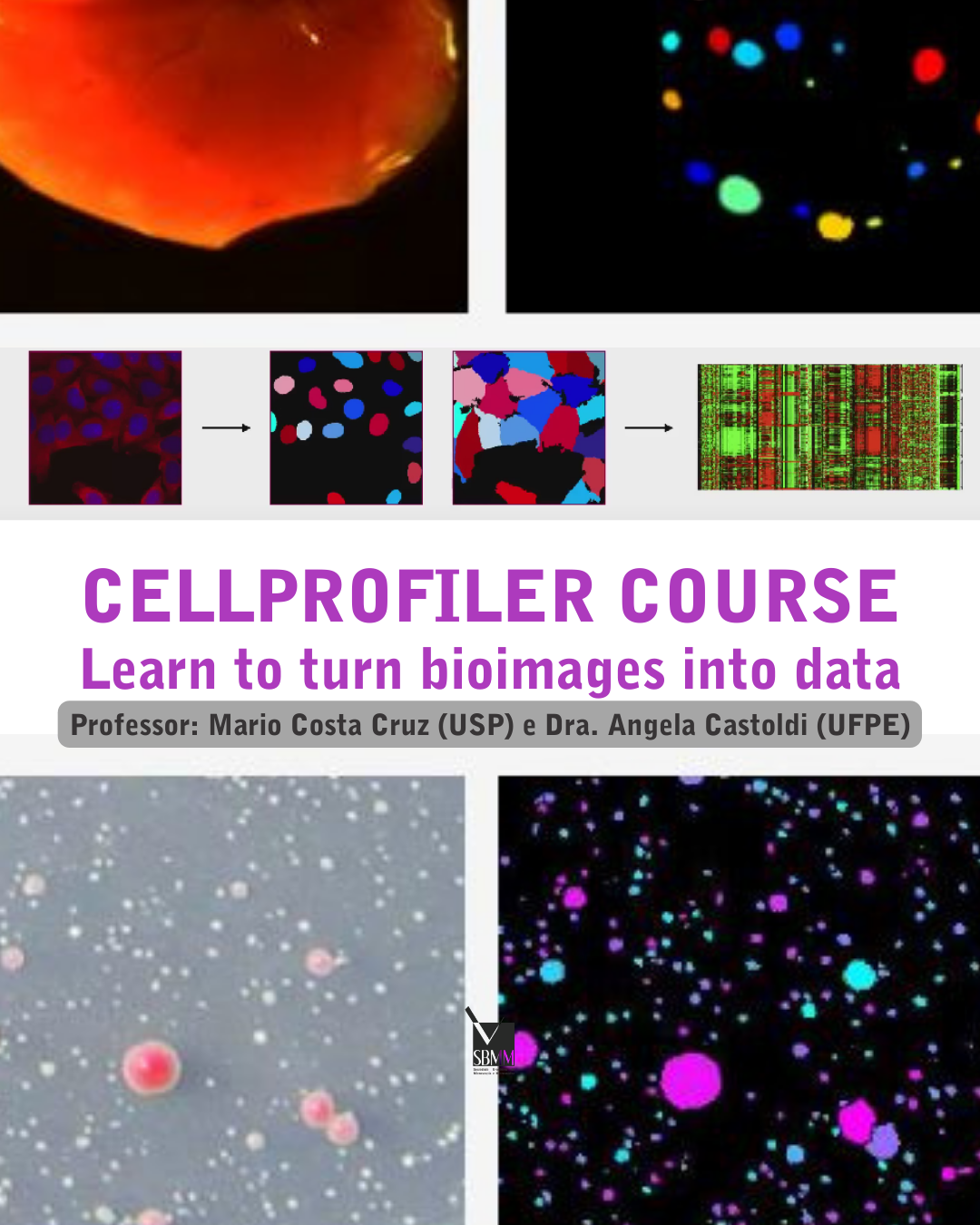
CellProfiler course - Learn to turn bioimages into data
Date: November 6th (Monday) between 9AM-12PM and 1:30PM-5PM.
Location: Laboratório de Imunopatologia Keizo Asami - Av. Prof. Moraes Rego, 1235 - Cidade Universitária, Recife - PE - CEP: 50670-901 - https://www.ufpe.br/ilika
Professors: Mario Costa Cruz (USP) and Dra. Angela Castoldi (UFPE)
Vacancies: 20
Requirement: Laptop (Mac, Linux or Windows)
Objective: Learn to turn bioimages into data using the free, open source software CellProfiler (https://cellprofiler.org/). This interactive workshop will go over the basics of creating a CellProfiler pipeline from start to finish within a focus on understanding how to adapt CellProfiler pipelines and module settings to new datasets. We will also demonstrate how to use the new CellProfiler plugins for deep learning-based segmentation with cellpose and stardist. The plugins runCellpose and runStardist make these algorithms more accessible and allow for their inclusion in existing CellProfiler workflows. The workshop is an introduction to CellProfiler and targeted to beginners with very little or no experience with the software.
Contact: mccruz@usp.br
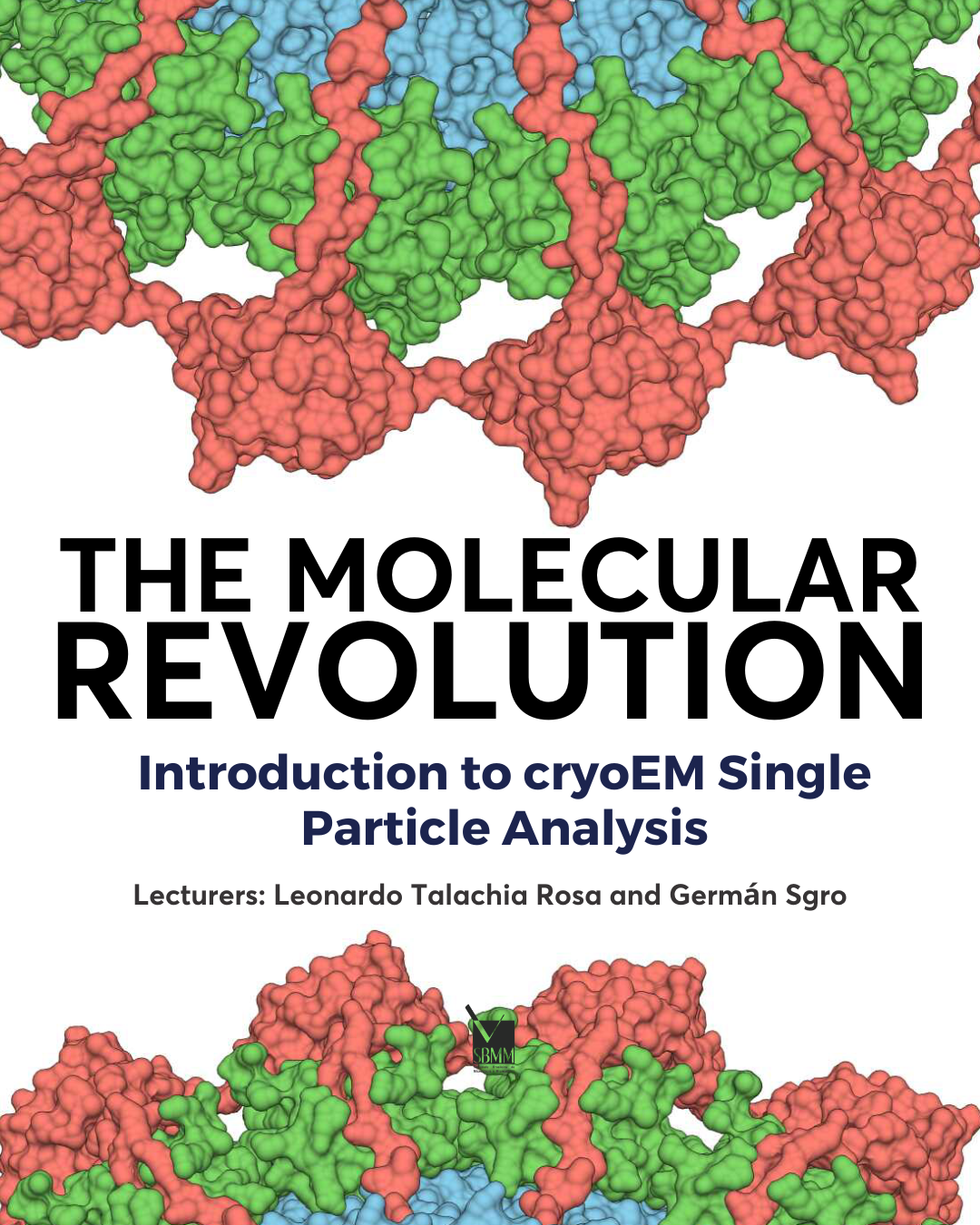
The molecular revolution - Introduction to cryoEM Single Particle Analysis
Date/hour: Monday (11/06 - 13:00-17:00 and 08:30 - 12:00).
Location: UFPE (more details soon)
Professors: Leonardo Talachia Rosa (USP) and Germán Sgro (USP)
Vacancies: 30
Objective: Cryo electron microscopy has revolutionized the field of structural biology in the last decade, as it allowed the near-atomic reconstruction of (multi)protein complexes in a state very close to the native state, leading to answers to biological questions hitherto unexplored. This mini-course aims to give an overview of this powerful technique, with a focus on characterization by Single Particle Analysis. Topics such as the potential applications and limitations of the technique, the necessary infrastructure, sample preparation and computational processing of the collected data will be addressed, so that the student can take the first steps towards the use of the technique in their research projects.
Contact: leotrosa@unicamp.br and ggsgro@usp.br
Abstract Submission
ABSTRACT CONTENT
Abstracts should be a condensed version of the final presentation and include all significant findings. Write the text so readers who are not specialists will be able to appreciate the purpose of the study and understand the procedures and conclusions. The text, entirely written in English, must include a short introduction and motivation for the study, including experimental procedures, main results, and conclusions. It is not necessary to divide the text into sub-sections.
INSTRUCTIONS FOR ABSTRACT PREPARATION (ORAL or POSTER)
1. Two Abstract can be submitted for each registration.
2. Extension/Format: A file for the entire abstract is .PDF
3. Your abstract should consist of exactly 2 pages, A4 format (201 x 297mm / 8.267 x 11.692in).
4. Submissions of less than 300 words will not be accepted.
1. The first page should have only text, no illustrations.
2. TITLE
Times New Roman, 14 point, boldface, alignment centered. Use initial capitals for each word and single line spacing if more than one line is needed for the title. Leave one line of space after the title.
3. AUTHOR'S NAMES
Times New Roman, 12 point, alignment centered. Type the authors' names in sequence, separated by commas. Each name must be followed by a superscript number as reference to each author's affiliation. Indicate the corresponding author with an asterisk after the superscript number. Use single line spacing if more than one line is needed for the author's names. Leave one line of space after author's names.
4. AUTHOR'S AFFILIATIONS
Times New Roman, 12 point, alignment left. List each author's affiliation in one line, preceded by the corresponding superscript number. Add the e-mail address only for the corresponding author. Leave one line of space after authors' affiliations.
5. TEXT
Times New Roman, 12 point, alignment justified (left and right). Write the text in just one paragraph. Use metric system (SIU) only.
6. REFERENCES
Indicate references in the text with Arabic numbers in square brackets, preferably at the end of the sentence, before the period. References must be included with the appropriate reference styles for proceedings abstracts [1], journal articles [2], and books [3]. Acknowledgments are included in the last reference, which is cited at the end of the last sentence of the text on the first page [4].
Collect references at the end of the text, on page 1 or page 2. For three or more authors, use first-named author's name plus “et al.” (See references 1 and 3 below). Do not use underlines on publication titles; use italics. Acknowledge sponsorship or support should be the last reference, corresponding to a numeral at the end of the text.
Examples:
[1] H.G. Hansma et al., Microsc. Microanal. 5 (Suppl. 2) (2002) 1012.
[2] M.A. Rodrigues, Thin Soli Films, 6 (2000) 1192.
[3] J.I. Goldstein et al., Scanning Electron Microscopy and X-ray Microanalysis, Plenum, New York, 1992.
[4] This research was supported by CNPq (Brazil).
1. The second page can comprise figures, figure captions and tables, as well as continuation of the text. Place table captions above tables at the left margin; place figure captions below figures.
2. LINE DRAWINGS, MICROGRAPHS AND PICTURES
All artwork must be electronically inserted into the Word document. Line art must be created either in a drawing program or scanned into a suitable format for importing into the document. Check that the width of lines and size of fonts allow the figure to be clear at its final size, if reduced. Be sure to include figure labels and scale markers as necessary.
3. TABLES
All tables must be electronically inserted into the Word document. Use the table-making functions of the Word processor to create the table with a horizontal rule at the top and bottom and below the column headings. Indicate units (in parentheses) in column headings as needed. Type text single-spaced within the table.
4. UPLOAD
All contributions must conform to the requirements above. Only Portable Document Format (PDF) will be accepted by the SSBMM electronic submission system. The final PDF file size should not exceed 5 MB.
5. ACCEPTANCE POLICY
Abstracts will be reviewed according to the following criteria: (a) relevance to a specific symposium, (b) scientific content and innovative proposals quality, (c) clarity of the text, and (d) compliance with the format. Abstracts not meeting the criteria above will not be accepted. Accepted abstracts must be presented by a registered author. Because major revisions may affect a symposium organizer's decision to accept your abstract, please review it carefully before submission. After submission, abstracts will not be accessible for corrections.
POSTER PRESENTATION
1. The poster sessions will be visited by evaluators, who will select the best papers from each session to receive an Honorable Mention at the closing ceremony.
2. The posters will be mounted on panels, where the works presented may occupy an area of 1.40 m high by 0.90 m wide.
3. Like abstracts, posters must be in English.
4. We remind you that, for publication in the book of abstracts and presentation of the work, at least one of the authors must be registered in the Congress.
5. Certificate of presentation work only for authors registered in the Congress.
LIST OF SYMPOSIA
MATERIALS SYMPOSIUM
- Scanning electron and Transmission electron microscopy and spectroscopy of nanostructures and engineering materials
- Advances in Scanning Probe Microscopy
- Advanced Imaging and Spectroscopy for materials applied to Sustainable processes
- Applications of focused ion beam instrumentation to materials
- 4D-STEM
- Microscopy in: Metallurgy and Mineralogy/Ceramics, metals, and minerals/Biomaterials
- In-situ Microscopy
- Radiation effects in materials
- Educational and capacity building in microscopy
- Measuring Cellular Properties using Atomic Force Microscopy
- Microscopy research applied to animal cells
- 3D Structures from In-cell structural biology to ultrastructure
- Cryo-EM of macromolecular assemblies
- Agriculture and Environment-Related research exploring microscopy tools
- Super-resolution microscopy
- Microscopy applied in Medical Science
- Microscopy imaging of pathogens, host and their interactions
- Live Imaging of Cells, Tissues and Organisms
- Microscopy for forensics
- Microscopy and Microanalysis in Industry
- Computational microscopy advances and perspectives
- Interface nano-bio